Graphical Abstract
Porous scaffolds and cell interfaces
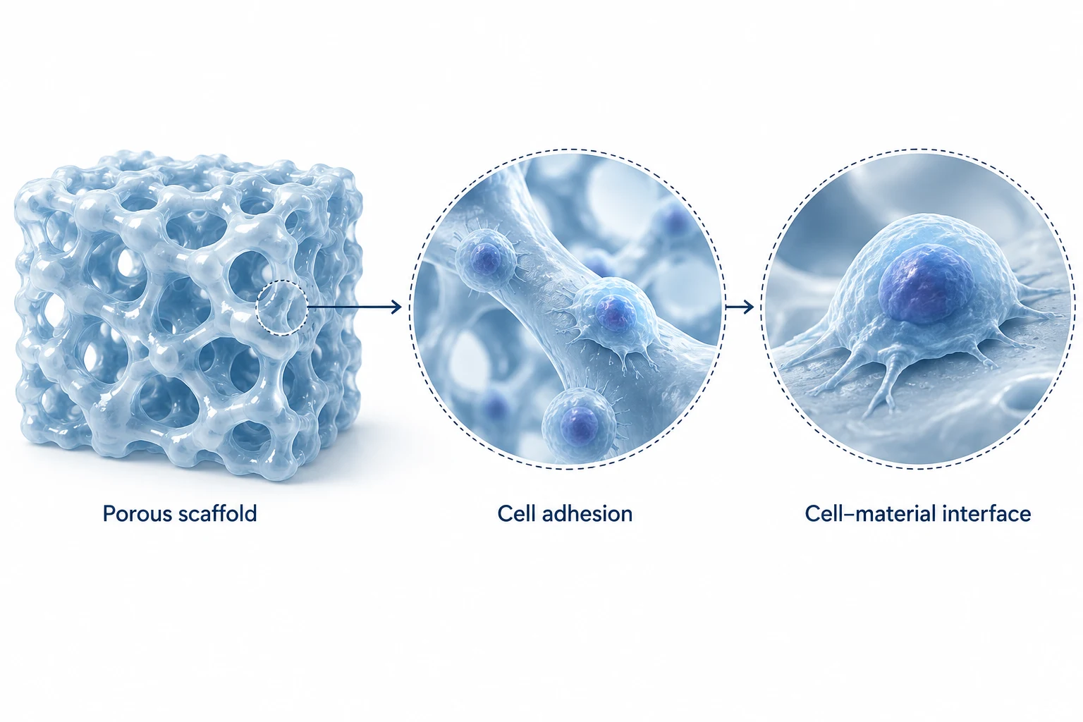
Three linked views move from porous architecture to cell adhesion and the cell–material interface, connecting material and cellular scales in a graphical abstract.
Read from the scaffold to the interface
The left view presents the overall porous scaffold. The middle view focuses on cells attached to a material surface, while the right view moves closer to a single cell and its contact with that surface. These views connect material and cellular scales for a graphical abstract about structure and interfaces.
What the arrows and enlarged views mean
Dashed circles identify areas of interest, and arrows guide the reader toward a closer view. They do not encode elapsed time, measured effects or a verified causal relationship. Cell shapes and material surfaces communicate spatial relationships; pore dimensions, scale and cell identity must be checked against the research material before adding specific labels.
Prepare a brief for a similar figure
Supply structural images or micrographs, cell names, the interface relationship to communicate and a short statement of the research finding. Separate experimental observations from structural relationships that the figure needs to explain. Decide which objects must remain visible in each view. Any dimensions or performance comparisons require their own measurement evidence.
Review before delivery
Review the structure and reading sequence first, followed by labels, color choices and legibility at the final display size. For submission, confirm the journal instructions, language, dimensions and file format. Agree editable files, detail views and alternate exports in the delivery checklist before production begins.